Initial Presentation to Cardiology
Koko, a young female spayed Sheltie Cross, was referred to our hospital after her primary care veterinarian detected a grade V/VI continous murmur during a routine check-up. While murmurs in younger dogs may be benign, these benign murmurs are usually up to grade III/IV and at the level of the heart base. Further investigation is always warranted in cases where grade III/IV murmurs do not disappear by 4-5 months of age, or in cases where louder murmurs, continuous murmurs or murmurs in other locations are detected, in order to rule out congenital diseases.
Further Investigation: Echocardiography and Doppler Findings
Our cardiology team performed a full cardiac work-up, including echocardiography and Doppler studies. The echocardiogram revealed that mild to moderate left ventricular enlargement and mild left atrial enlargement were present, with mild mitral regurgitation and a structurally normal mitral valve.
A Patent Ductus Arteriosus (PDA) was confirmed, showing left-to-right shunting with no evidence of pulmonary hypertension.
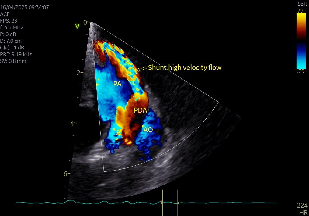
A short loop video showing the echocardiogram of the PDA.
Patent Ductus Arteriosus
Patent Ductus Arteriosus (PDA) is a congenital vascular anomaly where the ductus arteriosus, a foetal vessel that normally closes shortly after birth, remains patent. This results in persistent communication between the descending aorta and the main pulmonary artery, bypassing the systemic circulation’s usual path.
In postnatal circulation, the pressure in the aorta is significantly higher than in the pulmonary artery, leading to left-to-right shunting of blood through the PDA. This causes volume overload of the pulmonary circulation, left atrium, and left ventricle, as oxygenated blood is recirculated through the lungs rather than being distributed systemically.
It has been documented that if PDAs are left untreated, the patient would go on to develop congestive heart failure, and have a markedly reduced lifespan.
Early diagnosis and closure, either via surgical ligation or via transcatheter device occlusion, is essential to prevent irreversible cardiac changes.
Diagnosis and Surgical Decision
Despite her age, Koko had no major comorbidities and remained a good candidate for interventional correction. Our team, led by Dr Domingo Casamian-Sorrosal, Singapore’s only board-certified veterinary cardiologist, recommended PDA occlusion via a minimally invasive catheter-based technique – a first-of-its-kind procedure in Singapore.
PDA Occlusion: A Minimally Invasive Approach
Under fluoroscopic guidance, a catheter was advanced through the femoral artery and guided to the site of the PDA. An Amplatz Canine Duct Occluder (ACDO) device was deployed to seal the abnormal vessel. Fluoroscopy allowed real-time confirmation of the device’s position and successful closure of the ductus without the need for open-chest surgery.
Angiography using real-time fluoroscopy, showing how we advanced our catheter through the aorta to place the ACDO and occlude the PDA.
A loop video showing the closure of the PDA by the ACDO, with no blood flow occurring between the Pulmonary Artery (PA) and Aorta (AO).
Outcome and Recovery
Koko recovered smoothly post-op with no complications. At her 5-week recheck, echocardiography confirmed complete PDA closure, resolution of turbulent flow, and reverse remodelling of cardiac chambers. She was bright, active, and clinically well.
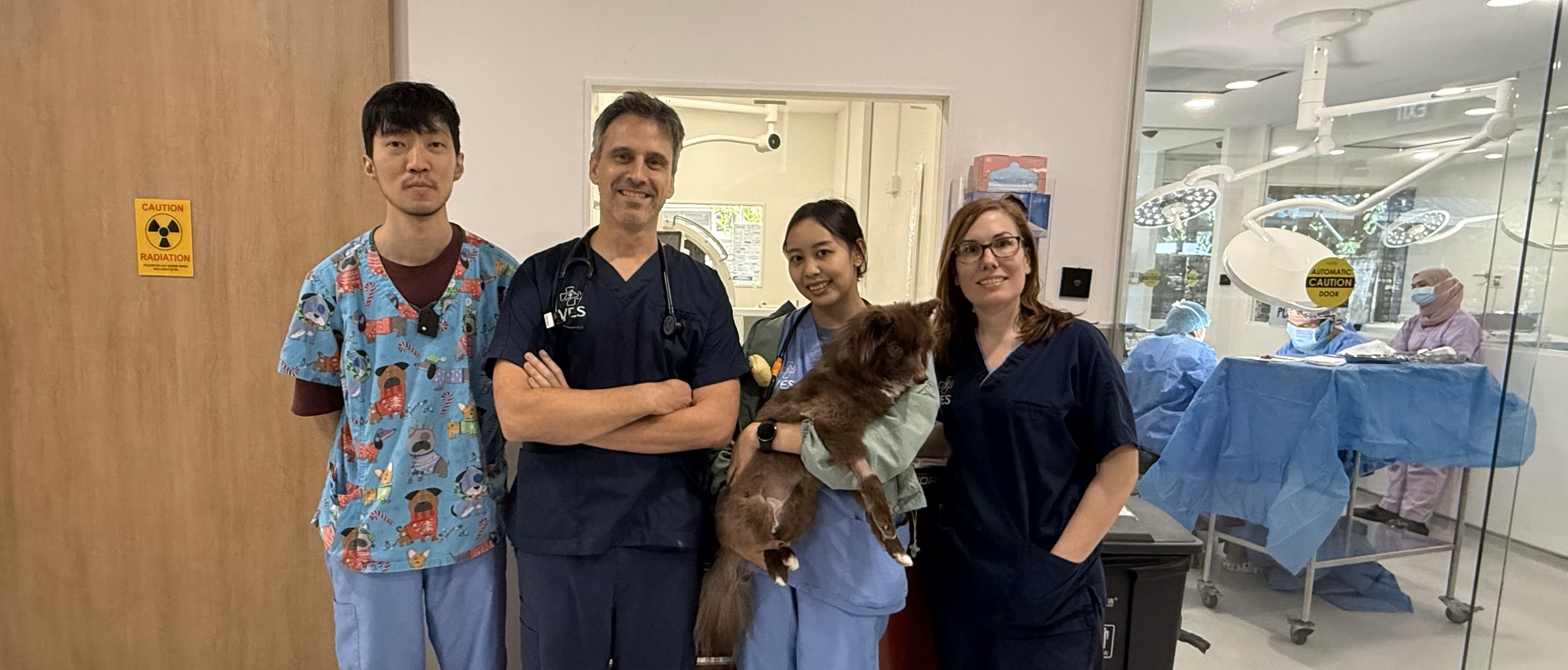
The success of this complex procedure was only made possible through the teamwork through our multidisciplinary specialist team, which included Dr Domingo our cardiologist, Dr Katherine our specialist anaesthesiologist, as well as the emergency and ICU team led by Dr Melissa and Dr Alex.
Why PDA Occlusion is Preferable to Traditional Surgery
Transcatheter PDA occlusion using devices such as the Amplatz Canine Duct Occluder (ACDO) offers several key advantages over traditional open-chest surgical ligation:
- Minimally invasive: The procedure is performed via a femoral artery catheter, avoiding thoracotomy and reducing surgical trauma.
- Reduced anaesthetic and recovery time: Patients typically recover faster with fewer complications and a shorter hospital stay.
While thoracic surgery remains an option in some cases (e.g. very small patients or abnormal duct anatomy), PDA occlusion has become the preferred method in suitable candidates due to its safety, efficacy, and minimally invasive nature.
A Reminder: Murmurs Matter in all Dogs!
Koko’s case is a reminder that not all heart murmurs in young dogs are innocent! A life-threatening congenital defect can be picked up like with Koko, and be repaired as soon as possible.
We encourage referring vets to send us any patients with unexplained murmurs for further evaluation. With early detection and advanced intervention, even congenital heart disease in adult dogs can be treated successfully.